
How can we help you?
Contact us at the Consultec WP office nearest to you or submit a business inquiry online.
CONTACT US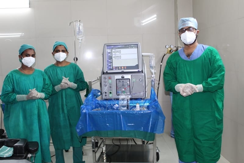
Vitreoretinal clinic
Ivision eye hospital have a vitroretinal clinic department to deal with all Vitreoretinal eye surgeries includes a group of procedures performed deep inside the eye's interior with lasers or conventional surgical instruments. As the name implies, this delicate surgery takes place where the gel-like vitreous and light-sensitive membrane (retina) are found. Also Ivision have folowing specilities in Vitreoretinal procedures
Fundus Fluorescein Angiography with digital imagin
Ivision medical team works with most advanced digital imaging equipment’s for Fundus Fluorescein Angiography, FA requires the use of a dedicated fundus camera equipped with excitation and barrier filters. Fluorescein dye is injected intravenously, usually through an antecubital vein with sufficient speed to produce high contrast images of the early phases of the angiogram. White light from a flash is passed through a blue excitation filter. Blue light (wavelength 465-490 nm) is then absorbed by unbound fluorescein molecules, and the molecules fluoresce, emitting light with a longer wavelength in the yellow-green spectrum (520-530nm). A barrier filter blocks any reflected light so that the images capture only light emitted from the fluorescein. Images are acquired immediately after injection and continue for ten minutes depending on the pathology being imaged. The images are recorded digitally a height resolution photograph. This detailed documentation help our doctors to precisely evaluate patient’s conditions like severity of the retinopathy etc.
B Scan imaging
Ivision eye hospital have B-scan Ultrasonography in our hospital, often we call it just B-scan or Brightness scan, offers two-dimensional cross-sectional view of the eye as well as the orbit. A B-scan is used on the outside of the closed eyelid to view the eye. This type of diagnostic tool is most helpful when there is difficulty examining the eye normally. There might be lid problems that make a routine examination difficult like edema or tarsorrhaphy or severe cataracts and keratoprosthesis. A B-scan can help accurately view other eye structures like the lens, choroid, sclera, vitreous and retina. A B-scan is helpful in diagnosing retinal detachment. A B-scan is often a tool combined with an A-scan to help determine various eye abnormalities.
Laser photo coagulation for diabetic retinopathy and vascular occlusions
For diabetic retinopathy ivision eye hospital uses Laser photo coagulation for diabetic retinopathy and vascular occlusions treatment technics Medicinal lasers are a standard source of light to produce retinal tissue photocoagulation to treat retinovascular disease. The Diabetic Retinopathy Study and the Early Treatment Diabetic Retinopathy Study were large randomized clinical trials that have shown beneficial effect of retinal laser photocoagulation in diabetic retinopathy and have dictated the standard of care for decades.
Laser indirect ophthalmoscopy
A Laser indirect ophthalmoscope constitutes a laser light attached to a headband, in addition to a small handheld lens. It provides a wider view of the inside of the eye. Furthermore, it allows a better view of the fundus of the eye, even if the lens is clouded by cataracts. An indirect ophthalmoscope can be either monocular or binocular. It is used for peripheral viewing of the retina to evaluate retina health. Ivision is equipped with most modern Laser indirect Ophthalmoscopes to provide the best available treatment in retina related problems.


©Microclouds 2018 . All rights reserved By Ivision Eye Hospitals.